What is a MRI?
MRI uses a powerful magnetic field, radio frequency pulses and a computer to produce detailed pictures of organs, soft tissues, bone and virtually all other internal structures of the human body.
The MRI consists of a large, cylinder-shaped magnet which is permanently on.
When you are in an MRI scanner, the strong magnetic field of the permanent magnet aligns the nuclei of the body´s atoms parallel to the magnetic field, like tiny magnets (normally the millions of nuclei all lie in random directions).
Then, short bursts of radio waves, 10 000 to 30 000 times stronger than the magnetic field of the earth, are sent from the scanner into your body.
This affects the body's atoms, forcing the nuclei into a different position.
When the burst of radio waves stops, the nuclei realign back into place. As they do so they emit radio signals. The nuclei in different tissues of the body realign at different speeds. Therefore, the signal emitted from different body tissues varies.
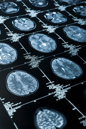
The scanner picks up these signals and a computer creates a picture based on the radio signals emitted from the body.
The images can then be examined on a computer monitor, transmitted electronically, printed or copied to a CD.
The human body consists mainly of water and most of the hydrogen atoms in the body are in water molecules. (water is made of hydrogen and oxygen – H20). In essence, MRI produces a map of hydrogen distribution in the body. The tissue that has the least hydrogen atoms (such as bones) turns out dark, while the tissue that has many hydrogen atoms (such as fatty tissue) looks much brighter.
A MRI scan can create detailed pictures of most parts of the body. So, it is useful where other tests (such as X-rays) do not supply adequate information required for a diagnosis. MRI is particularly useful for imaging the brain, spine, major joints and the interior structure of bones.
MRI does not use ionizing radiation (x-rays) and poses almost no risk to the average patient when appropriate safety guidelines are followed.
MRI Brain And Head
MRI is one of the few imaging tools that can see through bone (the skull) and deliver high quality pictures of the brain's anatomy and delicate soft tissue structures. MRI may be needed for patients with symptoms of a brain tumor, stroke, or infection (like meningitis). MRI also may be needed when cognitive and / or psychological symptoms suggest brain disease (like Alzheimer's or multiple sclerosis), or when developmental retardation suggests a birth defect.
MRI Spine
MRI is particularly useful for identifying and evaluating degenerated or herniated spinal discs and intervertebral joint disease, both frequent causes of severe lower back pain and sciatica (back pain radiating into a leg). It can also be used to determine the condition of nerve tissue within the spinal cord and explore other possible causes of back pain (compression fracture, infection or tumors, for example). It is a useful modality to help plan spinal surgical procedures, such as decompression of a pinched nerve or spinal fusion and monitor changes in the spine after an operation.
MRI Major Joints
In conjunction with conventional x-rays, MRI is usually the best choice for examining the body's major joints like the shoulder, wrist, hips, knees and ankles. MRI can provide clear images of the bone, cartilage, ligaments, and tendons that comprise a joint. MRI can be used to diagnose joint injuries due to sports, advancing age, or arthritis. A special form of MRI called an MR arthrogram involves the injection of contrast material into the joint so that the radiologist can better assess the relevant structures.
MRI Skeleton
The specific properties of MRI allow it to view the inside of bones. It can be used to detect bone cancer, inspect the marrow for leukemia and other diseases and examine subtle compression fractures of the spine due to injury, osteoporosis, infection or bone cancer.
MRI Rest Of Body
While CT and ultrasound satisfy most chest, abdominal, and general body imaging needs, MRI may be needed in certain circumstances to provide better pictures. MRI may also be used when repeated CT scanning is required and the effect of repeated x-ray exposure is a concern (i.e. the progress of some therapies, like liver cancer therapy, needs to be monitored with serial imaging).
How should I prepare for the MRI Scan?
Our MRI personnel will question you thoroughly prior to your examination to ensure that you can be imaged safely.
You may be asked to wear a gown during the exam or you may be allowed to wear your own clothing if it is loose-fitting and has no metal fasteners. Items like jewelry, watches, credit cards, hearing aids, pins, hairpins, metal zippers, removable dental work, pocketknives and eyeglasses, body piercings, and other accessories should be left at home if possible, or removed prior to the MRI scan.
Guidelines about eating and drinking before an MRI exam vary with the specific exam. Unless you are otherwise instructed, you may follow your regular daily routine and diet. Any prescribed medication may be taken.
Some MRI examinations may require the patient to receive an intravenous injection of contrast material. The contrast material most commonly used for an MRI exam, called Gadolinium, does not contain iodine and is less likely to cause side effects or an allergic reaction. Some conditions, such as severe kidney disease may prevent you from being given contrast material for an MRI. If there is a history of kidney disease, it may be necessary to perform a blood test before the examination to determine whether the kidneys are functioning adequately.
Women should always inform their doctor or radiographer if there is any possibility that they may be pregnant. Pregnant women should not have this exam unless the potential benefit from the MRI exam is assumed to outweigh the potential risks. Pregnant women should not receive injections of contrast material.
A person who is very large may not fit into the opening of the MRI machine (maximum weight 150 Kg).
Some people cannot have an MRI scan and should not enter the scanning area. If any of the following applies to you, please contact us before your appointment.
- Cardiac (heart) Pacemaker or implanted defibrillator.
- Cochlear (ear) implant.
- Some types of clips used on brain aneurysms.
- Some types of metal coils placed within blood vessels.
- Some types of artificial heart valves.
- Some types of implanted drug infusion ports.
- Any operation to your brain or heart.
- Any type of metal implant.
- Any injury to your eyes involving metal fragments.
- You are, or might be pregnant.
- Any kidney problems.
In general, metal objects used in orthopedic surgery pose no risk during MRI. However, a recently placed artificial joint (less than three months post – op) may require the use of another imaging procedure.
Patients with any shrapnel, bullets, or other pieces of metal which may be present in your eyes or body due to accidents may require an x-ray prior to an MRI.
Tooth fillings and braces usually are not affected by the magnetic field but they may distort images of the facial area or brain, so the radiographer should be aware of them.
Parents who accompany children into the scanning room also need to remove metal objects and notify the technologist of any medical or electronic devices they may have.
Initial assessment of spine, bones and joints is typically performed with x-rays. Please ensure that you bring them along for the MRI scan.
How is the MRI Scan performed?
The MRI unit has a large cylindrical-shaped tube surrounded by a circular magnet.
To begin, you will be comfortably positioned on a special table and then a device called a "coil" may be placed over or under you. These coils help the machine produce the most detailed and clear images of a specific body area. When you are comfortably positioned, the table will slide into the space into the center of the magnet.
If a contrast material will be used in the MRI exam, the radiologist or sister will insert an intravenous line into a vein in your hand or arm.
You will be moved into the magnet of the MRI unit and the radiographer will leave the room while the MRI examination is performed. You will usually be alone in the exam room during the MRI procedure. However, the radiographer will be able to see, hear and speak with you at all times using a two-way intercom. The patient is given an emergency bulb. This can be pressed at any time during the examination if the patient is distressed or anxious. An alarm is activated and the radiographer will stop the sequence and enter the room to reassure the patient.
MRI exams generally include multiple runs (sequences), typically lasting a few seconds to a few minutes at a time.
You will know when images are being recorded because you will hear tapping or thumping sounds when the coils that generate the radiofrequency pulses are activated. While the scanner is producing the images, you will hear a repetitive, rhythmic sound. This is easily alleviated by wearing earplugs or headphones (these will be offered to you prior to the examination).
It is important that you remain perfectly still while the images are being recorded. For some types of exams, you may be asked to hold your breath. You will be able to relax between imaging sequences, but will be asked to maintain your position without movement as much as possible.
Because of the length of time an MRI takes to complete, many young children and infants require sedation. A parent is allowed to stay in the room as long as they are also screened for safety in the magnetic environment.
In selected patients, conventional arthrography will be performed first. During this procedure contrast material will be injected into the joint of concern
(typically in the shoulder, hip or wrist) before MRI in order to image the joint structures in more detail.
When the exam is finished, the radiographer will help you off the table.
When the examination is completed, you may be asked to wait until the radiographer or radiologist checks the images in case additional images are needed. The radiologist will analyze and interpret your results and submit a comprehensive report to your referring doctor, who will discuss the findings with you.
The entire examination is usually completed within 60-90 minutes.
What will I experience during and after the MRI Scan?
Most MRI exams are painless.
However, some patients find it uncomfortable to remain still during MR imaging.
Others experience a sense of being closed-in (claustrophobia). Therefore, sedation can be arranged for those patients who anticipate anxiety, but fewer than one in 20 require it.
It is normal for the area of your body being imaged to feel slightly warm, but if it bothers you, notify the radiographer.
You may be offered or you may request earplugs to reduce the noise of the MRI scanner, which produces loud thumping and humming noises during imaging.
When the contrast material is injected, it is normal to feel coolness and a flushing sensation for a minute or two. Very rarely, patients may experience side effects or allergic symptoms in which case you must immediately notify the radiographer. A sister or radiologist will always be available for immediate assistance.
Mothers should not breastfeed their babies for 24-48 hours after contrast medium is given. However, it is safe to continue breastfeeding after receiving Gadolinium.
If you have not been sedated, no recovery period is necessary. You may resume your usual activities and normal diet immediately after the exam.
What will a MRI Scan cost?
Given all the advantages, doctors would undoubtedly prescribe MRI more frequently, but the MRI process is complex and costly. Generally, MRI is prescribed only when serious symptoms and / or negative results from other tests indicate a need. Many times other procedures are sufficient for the type of diagnosis needed.
All fee structures correspond to medical aid tariffs.
Most medical aids cover the costs of CT and MRI procedures. In some instances, a co-payment is required. There may also be a limit to the number of CT and MRI procedures allowable in a year. The medical aids require a referral letter from a specialist and prior authorization must be obtained. You can obtain this authorization yourself if you have all the relevant procedure and tariff codes or you can ask our qualified CT / MRI receptionists to assist you.
Private patients who pay immediately with cash or with Master or Visa Cards will be charged medical aid rates.
The account remains your responsibility.
In the event of non – payment by your medical aid, you will be held liable for the account and it should be paid within 30 days.