What are general X-Rays?
X-rays were discovered in 1895 by Wilhelm Conrad Rontgen and are the oldest and most frequently used form of medical imaging.
X-rays are a form of radiation like light or radio waves. X-rays pass through most objects, including the body. Once it is carefully aimed at the part of the body being examined, an x-ray machine produces a small burst of radiation that passes through the body, recording an image on photographic film or a special digital image recording plate.
Different parts of the body absorb the x-rays in varying degrees.
Dense bone absorbs much of the radiation while soft tissue, such as muscle, fat and organs, allow more of the x-rays to pass through them. As a result, bones appear white on the x-ray, soft tissue shows up in shades of gray and air appears black.
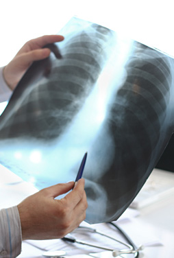
Chest And Sinuses
The chest and sinuses x-ray is performed to evaluate the lungs, heart and sinuses. A chest x-ray is typically the first imaging test used to help diagnose the cause of symptoms such as shortness of breath, a bad or persistent cough, chest pain, injury or fever. The examination is used to help diagnose or monitor treatment for conditions such as pneumonia, heart failure, emphysema and lung cancer.
Bone and Spine
A bone x-ray makes images of any bone in the body, including the hand, wrist, arm, elbow, shoulder, foot, ankle, leg, knee, thigh, hip, pelvis or spine. A bone x-ray is used to diagnose fractured bones or joint dislocation, demonstrate proper alignment and stabilization of bony fragments following treatment of a fracture, infection, arthritis, bone cancer and locate foreign objects in soft tissues around or in bones.
Pelvis And Lumbar Spine
Acute low back pain (LBP) with or without radiculopathy (nerve irritation caused by damage to the discs between the vertebrae) is one of the most common health problems and is a leading cause of disability for persons younger than age 45. Uncomplicated acute LBP or radiculopathy is a benign, self-limited condition that does not warrant any imaging studies and the vast majority of these patients are back to their usual activities within 30 days. The challenge for your doctor, therefore, is to distinguish that small segment that should be evaluated further because of suspicion of a more serious problem (symptoms getting worse and not resolving, unrelenting night pain or pain at rest, history or suspicion of cancer, fever above 38 degrees C for greater than 48 hours, osteoporosis, chronic oral steroids, immunosuppression,
serious accident or injury, clinical suspicion of ankylosing spondylitis, cauda equina syndrome, etc.).
Typically x-rays of the pelvis and lumbar spine may be requested if a patient has any of the "red flag conditions".
Patients over 50 years old with low back pain may warrant x-rays of the pelvis and lumbar spine due to the increased risk of malignancy, compression fractures etc.
Unnecessary or repeated x-rays should be avoided.
How should I prepare?
General x-rays require no special preparation.
You may be asked to remove some or all of your clothes and to wear a gown during the exam. You may also be asked to remove jewelry and any metal objects that might interfere with the x-ray images. These should preferably be left at home.
Women should always inform their doctor and x-ray personnel if there is any possibility that they may be pregnant. Many imaging tests are not performed during pregnancy so as not to expose the fetus to radiation. If an x-ray is necessary, precautions will be taken to minimize radiation exposure to the baby.
It is important to bring all previous, relevant x-rays with for comparison.
How is The Procedure Performed?
Special care is taken during x-ray examinations to use the lowest radiation dose possible while producing the best images for evaluation.
Chest And Sinuses
The equipment typically used for chest x-rays consists of a wall-mounted, box-like apparatus containing the x-ray film or a special plate that records the image digitally and an x-ray producing tube, that is usually positioned about two meter away.
Typically, two views of the chest are taken, one from the back and the other from the side of the body. The radiographer will position the patient with hands on hips and chest pressed against the image plate. For the second view, the patient's side is against the image plate with arms elevated. Patients who cannot stand may be positioned lying down on a table for chest x-rays.
You must hold very still and may be asked to keep from breathing for a few seconds while the x-ray picture is taken to reduce the possibility of a blurred image.
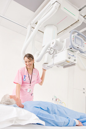
Bones, Joints And Spine
The equipment typically used for bone x-rays consists of an x-ray tube suspended over a table on which the patient lies. A drawer under the table holds the x-ray film or image recording plate. Sometimes the x-ray is taken with the patient standing upright, as in the case of knee x-rays.
The radiographer positions the patient on the x-ray table and places the x-ray film holder or digital recording plate under the table in the area of the body being imaged. When necessary, sandbags, pillows or other positioning devices will be used to help you maintain the proper position. A lead apron may be placed over your pelvic area or breasts when feasible to protect from radiation.
You must hold very still and may be asked to keep from breathing for a few seconds while the x-ray picture is taken to reduce the possibility of a blurred image.
Two or three images from different angles will typically be taken.
An x-ray may also be taken of the unaffected limb of a child for comparison purposes.
When the examination is complete, you will be asked to wait until the radiologist determines that all the necessary images have been obtained.
X-ray examinations are very useful, but they have limitations. Because some conditions of the chest cannot be detected on a conventional chest x-ray image, this examination cannot necessarily rule out all problems in the chest. For example, small cancers or a blood clot (pulmonary embolism) may not show up on a chest x-ray. Likewise, while x-ray images are among the clearest, most detailed views of bone, they provide little information about muscles, tendons or joints.
The Radiologist may suggest further imaging studies to clarify the results of the x-rays or to look for abnormalities not visible on the general x-rays.
Follow-up examinations are often necessary, and your doctor will explain the exact reason why another exam is requested. A follow-up examination may be necessary so that any change in a known abnormality can be detected over time. Follow-up examinations are sometimes the best way to see if treatment is working or if an abnormality remains stable over time.
The Radiologist will analyze the images and prepare a signed report for your referring doctor, who will discuss the results with you.
General x-rays are usually completed within 30 – 60 minutes.
What will I Experience During and After The Procedure?
An x-ray examination itself is a painless procedure.
Individuals with arthritis or injuries may have discomfort trying to stay still during the examination. The radiographer will assist you in finding the most comfortable position possible while still ensuring good diagnostic image quality.
What will it Cost?
Most medical aids will cover the cost of general x–rays provided that you have not exceeded your annual imaging limit.
Pre-authorization is usually not required by medical aids, but please consult with your medical aid if you are uncertain.
Private patients who pay immediately with cash or with Master or Visa Cards will be charged medical aid rates.
The account remains your responsibility.
In the event of non-payment by your medical aid, you will be held liable for the account and it should be paid within 30 days.